Coming soon!

About the Species
This specimen was made available to the University of Texas High-Resolution X-ray CT Facility for scanning by Dr. James Hanken of Harvard University. Funding for scanning was provided by an NSF grant (DEB-0345885) to Dr. Hanken, and funding for image processing was provided by an NSF Digital Libraries Initiative grant to Dr. Timothy Rowe of The University of Texas at Austin.
About this Specimen
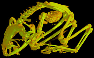
This specimen was scanned by Matthew Colbert on 31 October 2007 along the coronal axis for a total of 675 slices. Each 1024 x 1024 pixel slice is 0.05353 mm thick, with an interslice spacing of 0.05353 mm and a field of reconstruction of 25 mm.
About the
Scan
Literature
& Links
Front page image.
|  |
Additional
Imagery
|


 )


)