This page serves supplemental imagery for a paper entitled The Cephalic Osteoderms of Varanus komodoensis as Revealed by High-Resolution X-ray Computed Tomography by J.A. Maisano, T.J. LaDuc, C.J. Bell and D. Barber (In press, The Anatomical Record). The abstract is as follows:
Osteoderms constitute a morphological system that plays an important role in squamate systematics. However, their study has always been difficult due to their isolated occurrence in the skin, among the first organs to be removed during the skeletonization process. High-resolution X-ray computed tomography (HRXCT) offers a nondestructive means of visualizing osteoderms both in their natural relationship to each other and to the underlying cranial bones. While it is often stated that Varanus komodoensis has a chain mail of osteoderms, this morphological system was never described in this taxon. Further, given its size, it might be expected that V. komodoensis would present the extreme of osteoderm development in extant varanids, a group that tends to have weakly-developed osteoderms or none at all. Indeed, our HRXCT scan of a 19-year-old captive individual reveals an elaborate mesh of cephalic osteoderms that are incredibly numerous and morphologically diverse. We describe this skeletal system and compare it to the cephalic osteoderms in other varanoids.
Click here to download the original HRXCT data (3.4 Gb).

About the Species
This specimen, a male, was born at the National Zoological Park on 2 September 1993 and died 4 March 2013 at the Fort Worth Zoo. He lived for 19 years and 6 months, and was part of the first clutch of Komodo dragons to be produced in the United States.
The specimen was donated to the Texas Natural History Collections by Diane Barber, Curator of Ectotherms at the Fort Worth Zoo. He was made available to The University of Texas High-Resolution X-ray CT Facility for scanning by Dr. Jessie Maisano and Dr. Travis LaDuc of The University of Texas. Funding for scanning was provided by NSF grant EAR-1258878 to R. Ketcham, W. Carlson and T. Rowe.
About this Specimen
The specimen was scanned by David Edey on 2 April 2015 along the coronal axis for a total of 1816 slices. Voxel size is 0.1626 mm.
Click here to download the original HRXCT data (3.4 Gb).
About the
Scan
Literature
Erickson, G.M., de Ricqles, A., de Buffrénil, V., Molnar, R.E., and Bayless, M.K. 2003. Vermiform bones and the evolution of gigantism in Megalania how a reptilian fox became a lion. Journal of Vertebrate Paleontology, 34, 966-970.
Links
Varanus komodoensis page on Wikipedia
V. komodoensis page on The Reptile Database
Literature
& Links
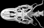
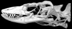
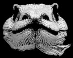
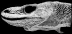
Three-dimensional volumetric renderings of the skull with the osteoderms digitally removed, and of the osteoderms with the skull digitally removed. All are less than 5mb.
Additional Imagery
 |


 )