|
|
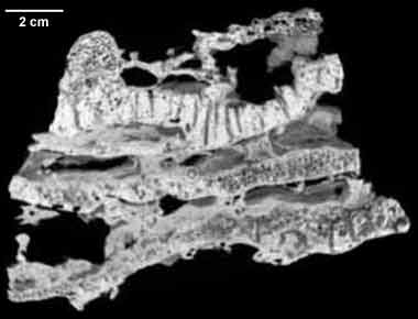
This specimen is a composite of Agaricia sp., Cliona sp. and Siphonodictyon sp. borings, and Halimeda sp. flakes and worm tubes. See "About this Specimen" for more information. 
About the Species
The reef rock in this study is from Discovery Bay, 1 km north of Jamaica. Discovery Bay is the site of a fringing reef complex with shallow and deep forereef components. The prograding shallow component ends with an abrupt drop-off at 55 meters depth. The deep forereef cliff then continues vertically to approximately 122 meters below sea level. The deep forereef has a corrugated appearance, the result of lateral reef growth (promontories) alternating with recessed drainage channels (reentrants). The latter are sparsely colonized due to high sedimentation rates. Promontories are dominated by the scleractinian corals Agaricia and Montastrea cavernosa and the calcareous green algae Halimeda. Hermatypic corals grow where light is available as symbiotic, photosynthesizing algae (zooxanthellae) live within their tissues. As light penetration diminishes with depth, these corals grow upward and outward and forms tend to be flattened to increase the amount of surface area exposed to light. Corals tilt seaward, preventing sediment accumulation on flat coral surfaces.
In July 1970, a team of scientists used explosives in the deep forereef and recovered the resulting reef talus, including the sample used in this study. The sample is characteristic of the deep forereef. Three layers of Agaricia plates are obvious though it is unclear if more than one colony is present. Lateral bands indicate growth direction. Encrusting sponges are obvious on the exterior, as are the effects of two boring sponges. Smaller borings are the result of Cliona, whereas Siphonodictyon produces larger holes. Halimeda flakes and worm tubes are also present. Micritic ooze and internal sediment can also be distinguished. Geopetals are present, indicating angle of in situ orientation.
The reef talus is irregularly shaped. Maximum dimensions are 10x16x12 cm. Dr. Lynton Land, professor emeritus of the Department of Geological Sciences of The University of Texas at Austin, provided the sample used in this study.
About this Specimen
Serial and thin sectioning are commonly used in the study of fossil and Recent reef ecology and sedimentology. Serial sectioning provides unique information to reef studies and is necessary for petrographical analysis of sediment, cement and organisms. However, serial sectioning also destroys valuable information. Sediments wash out, pieces break off and centimeters of rock are pulverized in the cutting process. In a CT scan, this information, important to the understanding of ecological and sedimentary processes, can be viewed easily and quantified in three dimensions. Digital images therefore preserve depositional relationships, saving information which can be invoked in concert with slab and thin section analysis. Digital images can be imported into programs such as NIH Image where aspects of coral biology (e.g., polyp size and growth rings) and ecology (e.g., bioerosion rates) can be quantified.
Despite these advantages, scanning of invertebrates has thus far been generally unsuccessful. The similarities in the effective density of the fossil, and it's encasing matrix, is not conducive to X-ray resolution of structures.
For instance, a test scan of a rock from the Capitan Reef complex of West Texas yielded an image that shows only a gray grainy matrix. The X-ray's response is the same for both the carbonate skeletal material of the organisms, and the carbonate sediments and cement.
Attempts to examine the internal structure of a fossilized echinoderm were similarly unusable. Digital scan results of a hermatypic coral (Monastrea cavernosa), however, were excellent, due to the effective density differences between the coral and the pore space of the polyp chambers.
This website shows the initial results of CT scanning on a Recent coral reef sample. The scan successfully shows coral framework, porosity, and internal sediments.
For scanning, the rock was oriented so that slice planes closely approximate the growth direction of the corals. The scanner was calibrated to subtract out detector noise and to define the samples center of rotation and the distances between x-ray source, sample and detector. X-ray detectors were restricted to 0.4 mm, the selected slice thickness for this sample. High energy x-rays were produced to ensure sample penetration. After a few test scans, 250 slices were taken over a 10 cm section of the rock.
Images must be processed to optimize data and remove the artifacts inherent to the current CT technology. Manipulating energy and resolution at the scanning level can decrease the prominence of these artifacts. Processing in NIH Image 1.6 and Adobe Photoshop can eliminate or reduce some of them, though artifacts are often impossible to delete without destroying image data. Ring artifacts are prominent in this data set, and are probably the result of temperature variations in the detector.
In NIH Image, the individual image files were stacked and cropped to reduce file size. Files were then unstacked and imported to Adobe Photoshop 4.0. In this program, a predetermined set of processing actions can be applied to an entire set of files, or batched. Batching ensures that the same changes apply to all the image files. Batch processing included adjusting levels to improve differentiation of organisms and sediments and deleting the container which the rock sat in during scanning.
The processed images were then imported back to NIH Image, where the stack of images was resliced in both horizontal and vertical planes. To make certain that resliced images are of the same scale as the original image, the scale of the image size on the screen was compared to the actual sample dimensions. This proportion is used to determine the thickness of the slice, in pixels, in other planes. In this sample, 180 mm distance on the rock sample is equal to 512 pixels on the screen. Therefore, 2.84 pixels equals one millimeter on the actual specimen. Since the original CT slices are 0.4 mm thick, new slices created in NIH image must each be 1.13 pixels thick (2.84 pixels/mm multiplied by 0.4 millimeters). Resulting slices will be at the same scale of the original slices and therefore, of the actual sample.
The slice planes are named after the directional components- XY, YZ and XZ. The XY and XZ planes are vertical and run perpendicular to each other. The YZ plane is horizontal and runs perpendicular to the vertical planes.
Funding for this project was provided by Dr. Brenda Kirkland-George and Dr. Rich Ketcham performed the CT scans in the fall of 1997.
About the
Scan
Literature
Aronson, R. B. and W.F. Precht. 1997. Stasis, biological disturbance, and community structure of a Holocene coral reef. Paleobiology 23:326-346.
Brusca, R.C. and G.J. Brusca. 1990. Invertebrates. Sunderland, MA: Sinauer.
Chazottes, V., T. Le Campion-Alsumard, and M. Peyrot-Clausade. 1995. Bioerosion rates on coral reefs: interactions between macroborers, microborers and grazer (Moorea, French Polynesia). Palaeogeography, Palaeoclimatology, Palaeoecology 113:189-198.
Conway Morris, S., J.D. George, R. Gibson, and H.M. Platt, eds. 1985. The Origin and Relationships of Lower Invertebrates. Oxford: Clarendon.
Fagerstrom, J.A. 1987. The Evolution of Reef Communities. New York: Wiley.
Humann, P. 1993. Reef Coral Identification. Jacksonville: Paramount.
Jones, O.A. and R. Endean. Biology and Geology of Coral Reefs: Volumes I and II. New York: Academic.
Land, L.S. 1977. The fate of reef-derived sediment on the North Jamaican island slope. Marine Geology 29:55-71.
Land, L.S. and C.H. Moore, Jr. 1977. Deep forereef and Upper Island slope, North Jamaica. Studies in Geology 4:53-65.
Veron, J.E.N. 1995. Corals in Space and Time. Ithaca: Cornell University.
Veron, J.E.N., J.M. Odorico, C.A. Chen, and D.I. Miller. 1996. "Reassessing evolutionary relationships of scleractinian corals." Coral Reefs 15:1-9.
Wood, Elizabeth. 1983. Corals of the World. Neptune City, NJ: T.F.H.
Links
General Information about corals and reefs
Reef health and monitoring
University and Research Stations
Literature
& Links
None available.
Additional
Imagery
|
|