|
|
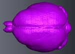
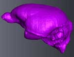
The imagery on this page was the basis for a paper entitled Cranial anatomy of the Duchesnean primate Rooneyia viejaensis: new insights from high
resolution computed tomography by E.C. Kirk, P. Daghighi, T.E. Macrini, B-A.S. Bhullar, and T.B. Rowe (2014, Journal of Human Evolution, 74, 82-95). The abstract is as follows:
Rooneyia viejaensis is a North American Eocene primate of uncertain phylogenetic affinities.
Although the external cranial anatomy of Rooneyia is well studied, various authors have suggested that
Rooneyia is a stem haplorhine, stem strepsirrhine, stem tarsiiform, or stem anthropoid. Here we
describe the internal cranial anatomy of the Rooneyia holotype based on micro-computed tomography
and discuss the phylogenetic implications of this anatomy. Precise measurements of the natural
endocast filling the braincase of the Rooneyia holotype reveal that the genus had a relative brain size
comparable to some living callitrichines and strepsirrhines. Rooneyia was thus probably more
encephalized than any other known omomyiform, adapiform, or plesiadapiform. Relative olfactory
bulb size in Rooneyia was most comparable to some living strepsirrhines and the stem anthropoid
Parapithecus. The nasal fossa of Rooneyia resembled that of living strepsirrhines in retaining a rostrocaudally
oriented nasolacrimal canal, four ethmoturbinals, and an olfactory recess separated from the
nasopharyngeal meatus by a transverse lamina. The ear region of Rooneyia is characterized by large
and complete canals for both the stapedial and promontory branches of the internal carotid artery.
Rooneyia also retains a patent parotic fissure and thus had an extrabullar origin of the stapedius
muscle. In most of these respects, Rooneyia exhibits the condition that is presumed to be primitive for
crown primates and lacks a number of key crown haplorhine synapomorphies (e.g., a dorso-ventrally
oriented nasolacrimal canal, loss of the olfactory recess, loss of ethmoturbinals 3-4, involution of the
stapedial artery). These data are consistent with the hypothesis that Rooneyia is an advanced stem
primate or a basal crown primate but are inconsistent with prior suggestions that Rooneyia is a crown
haplorhine.
See 'About the Scan' to download original high-resolution X-ray CT data sets. See 'Additional Imagery' for animations of the cranial endocast.

About the Species
Rooneyia viejaensis is a Late Eocene primate from the Sierra Vieja of West Texas. The species is known only from the type specimen (TMM 40688-7), which was recovered in 1964 from the Duchesnean Rifle Range Hollow locality in the Chambers Tuff Formation (Wilson, 1966; Wilson, 1986; Robinson et al., 2004). The Rooneyia holotype is a nearly undistorted cranium missing only the premaxillae, postorbital bars, zygomatic arches, and portions of the neurocranium. The specimen also preserves a complete and undistorted natural endocast of the braincase (Hofer and Wilson, 1967).
Funding for scanning of the whole skull was provided by a National Science Foundation Digital Libraries Initiative grant to Dr. Timothy Rowe of The University of Texas at Austin. Funding for close-up scanning of the ear was provided by Dr. Chris Kirk of The University of Texas at Austin. Additional funding for image processing was provided by the Jackson School of Geosciences. TMM 40688-7 is housed at the Jackson School of Geosciences Vertebrate Paleontology Laboratory.
About this Specimen
The whole specimen was scanned by Matthew Colbert on 5 September 2003 along the coronal axis for a total of 585 slices. In-plane resolution (X and Y) was 0.0391 mm, and interslice spacing (Z) was 0.0867 mm. This data set can be downloaded here (694 Mb).
A close-up scan of the right ear was done by Matthew Colbert on 8 June 2010 along the coronal axis for a total of 453 slices. In-plane resolution (X and Y) was 0.0117 mm, and interslice spacing (Z) was 0.0164 mm. This data set can be downloaded here (689 Mb).
The skull of Microcebus rufus, used for comparative purposes, was scanned by Matthew Colbert on 20 August 2007 along the coronal axis for a total of 670 slices. In-plane resolution (X and Y) was 0.0488 mm, and interslice spacing (Z) was 0.0535 mm. A cropped version of this data set can be downloaded here (42 Mb).
About the
Scan
Literature
Hofer, H.O., Wilson, J.A., 1967. An endocranial cast of an early Oligocene primate. Folia Primatol. 5, 148-152.
Kirk, E.C., Daghighi, P., Macrini, T.E., Bhullar, B-A.S., Rowe, T.B. In Review. Cranial anatomy of the Duchesnean primate Rooneyia viejaensis: new insights from high resolution computed tomography. Journal of Human Evolution.
Robinson, P., Gunnell, G.F., Walsh, S.L., Clyde, W.C., Storer, J.E., Stucky, R.K., Froehlich, D.J., Ferrusquia-Villafranca, I., McKenna, M.C., 2004. Wasatchian through Duchesnean biochronology. In: Woodburne, M.O. (Ed.), Late Cretaceous and Cenozoic Mammals of North America. Columbia University Press, New York, pp. 106-155.
Wilson, J.A. 1966. A new primate from the earliest Oligocene, West Texas, preliminary report. Folia Primatologica. 4, 227-248.
Wilson, J.A. 1986. Stratigraphic occurrence and correlation of early Tertiary vertebrate faunas, Trans-Pecos Texas: Agua Fria-Green Valley Areas. Journal of Vertebrate Paleontology. 6, 350-373.
Szalay, F.S., Wilson, J.A., 1976. Basicranial morphology of the early Tertiary tarsiiform Rooneyia from Texas. Folia Primatologica. 25, 288-293.
Links
More primates on the eskeletons website (The University of Texas at Austin)
Literature
& Links
Additional Imagery
 |
|


 ,
,  )