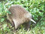
Zaglossus bartoni, the long-nosed echidna, is one of the five extant members of Monotremata, the others being Z. attenboroughi, Z. bruijni, Tachyglossus aculeatus and Ornithorhynchus anatinus. Monotremes, or egg-laying mammals, are named for the single common opening for the urogenital and digestive systems. Most phylogenetic analyses based on morphological and molecular data (e.g., Phillips and Penny, 2003) place Monotremata as the sister taxon to Theria (Placentalia + Marsupialia) among major clades of living mammals. An alternative hypothesis based on some molecular and morphological data places Monotremata as the sister taxon to Marsupialia (Gregory, 1947; Penny and Hasegawa, 1997). |
|
Zaglossus is found in the central cordillera and the mountains of the Vogelkop of New Guinea (Griffiths et al., 1991:87). Zaglossus is currently listed on appendix II of CITES. The fossil record of echidnas is poor, extending back only to the Pleistocene of Australia and New Guinea (Murray, 1978; Griffiths et al., 1991). The long-nosed echidna lives in humid montane forests and alpine meadows. Zaglossus is mainly nocturnal and exists on a diet primarily of earthworms (Griffiths et al., 1991; Nowak, 1991).
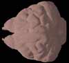
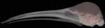
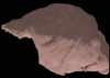
Echidnas, also known as spiny anteaters, are covered by hollow spines that are essentially modified hairs. The body is rounded and dorsoventrally compressed, ending in a short tail. The legs are short and stout; each ending in feet with five digits terminating in elongate claws. The most prominent feature on the head is the elongate, hairless snout. The mouth is toothless and contains a long, sticky tongue.
Zaglossus is larger than Tachyglossus and has a significantly longer snout, making up two-thirds of the entire length of the head (Nowak, 1991). The spines of Zaglossus are shorter and less numerous than those of Tachyglossus (Nowak, 1991).
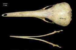
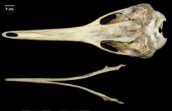
Similar to Tachyglossus, the skull of Zaglossus is characterized by an elongated, rounded snout and a laterally bulging braincase. The palate extends back to the ears. The ectotympanic is oriented horizontally and the external auditory meatus is ventrally directed. The lower jaw is extremely reduced and has poorly developed coronoid and angular processes. Inside the skull, the most prominent features are the turbinates. They extend far posteriorly in the skull, underlying the olfactory and cerebral cavities of the braincase. Differences between the skulls of Zaglossus and Tachyglossus are discussed more thoroughly in Griffiths et al. (1991).

Literature
Allen, G. M. 1912. Zaglossus. Memoirs of the Museum of Comparative Zoology at Harvard College 40:253-307.
Augee, M. L. (ed.) 1978. Monotreme biology. The Australian Zoologist 20, part 1.
Augee, M. L. (ed.) 1992. Platypus and echidnas. Royal Zoological Society of New South Wales, Mosman, 296 pp.
Flannery, T. F., and C. P. Groves. 1998. A revision of the genus Zaglossus (Monotremata, Tachyglossidae), with description of new species and subspecies. Mammalia 62:367-396.
Gregory, W. K. 1947. The monotremes and the palimpsest theory. Bulletin of the American Museum of Natural History 88:5-52.
Griffiths, M. 1968. Echidnas. Pergamon Press, New York, 282 pp.
Griffiths, M. 1978. The biology of the monotremes. Academic Press, New York, 367 pp.
Griffiths, M., R. T. Wells, and D. J. Barrie. 1991. Observations on the skulls of fossil and extant echidnas (Monotremata: Tachyglossidae). Australian Mammalogy 14:87-101.
Lyne, G. 1967. Marsupials and monotremes of Australia. Taplinger Publishing Company, New York, 72 pp.
Murray, P. F. 1978. Late Cenozoic monotreme anteaters. Pp. 29-55 in: Monotreme biology. The Australian Zoologist 20, part 1.
Nowak, R. M. 1991. Walkers Mammals of the World. Volume 1. Fifth edition. The Johns Hopkins University Press, Baltimore.
Penny, D., and M. Hasegawa. 1997. The platypus put in its place. Nature 387:549-550.
Phillips, M. J., and D. Penny. 2003. The root of the mammalian tree inferred from whole mitochondrial genomes. Molecular Phylogenetics and Evolution 28:171-185.
Watson, D. M. S. 1916. The monotreme skull: a contribution to mammalian morphogenesis. Philosophical Transactions of the Royal Society of London B 207:311-374.
Links
Zaglossus bruijni on the Animal Diversity Web (Univ. of Michigan Museum of Zoology).


 )